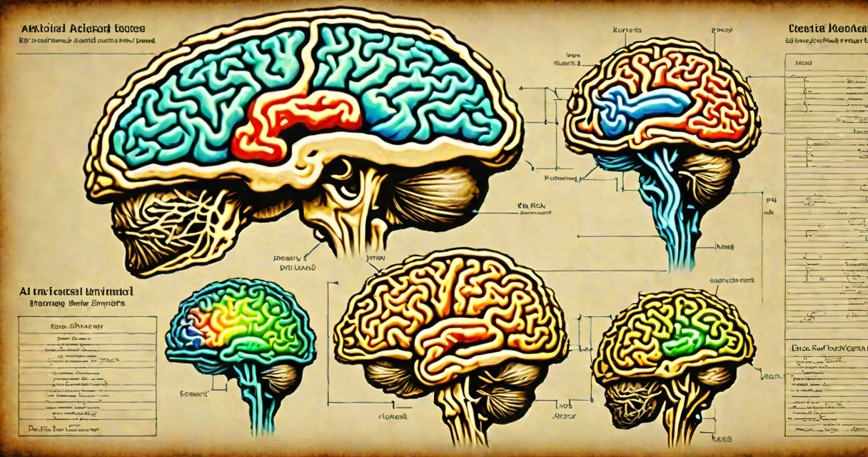
AI in medical imaging uses machine learning models to analyze X-rays, MRIs, CT scans, and ultrasounds to detect diseases such as cancer, stroke, and fractures with speed and consistency exceeding human radiologists in specific tasks.
These systems process medical images by identifying patterns in pixel data that correlate with known pathologies. They're trained on thousands of labeled scans-where experts have annotated findings like nodules in lungs or lesions in brain tissue. Once trained, the AI can flag potential issues in new scans within seconds, often catching subtle changes invisible to the naked eye.
For example, Google Health's AI model reduced false negatives in breast cancer detection on mammograms by 9.4% compared to radiologists alone, according to a 2020 study published in Nature. This improvement came from analyzing dense breast tissue, where early cancers are harder to spot.
AI in medical imaging relies on deep learning algorithms, particularly convolutional neural networks (CNNs), which mimic how the visual cortex processes shapes and textures. These networks scan images layer by layer, extracting features like edges, contrast gradients, and spatial relationships.
During training, the model adjusts internal weights based on feedback from correct diagnoses. After sufficient iterations, it learns to distinguish between healthy tissue and abnormal growths. During deployment, it outputs a probability score for each suspected condition-often displayed as heatmaps overlaid on the original image.
A clinical workflow might involve a technician uploading a chest CT scan. The AI flags a 6mm nodule in the lower lobe. A radiologist then reviews the area, confirming the finding. The AI didn't replace the expert-it accelerated attention to high-risk areas.
Different AI tools serve distinct roles across imaging modalities:
| Modality | Common AI Use Cases | Accuracy Benchmark (from peer-reviewed studies) |
|---|---|---|
| MRI (brain) | Stroke detection, multiple sclerosis lesion tracking | 92% sensitivity in detecting acute ischemic stroke (JAMA Neurology, 2021) |
| CT (chest) | Lung nodule classification, pneumonia severity scoring | 94% specificity in distinguishing benign from malignant nodules (Radiology, 2022) |
| X-ray (chest) | Tuberculosis screening, pneumothorax detection | 88% accuracy in detecting collapsed lungs (Nature Digital Medicine, 2023) |
| Ultrasound (abdomen) | Fatty liver assessment, gallstone identification | 91% agreement with expert sonographers (Ultrasound in Medicine & Biology, 2022) |
Some AI tools focus on quantification-measuring tumor volume over time. Others prioritize speed, processing hundreds of scans per hour. A few systems integrate with electronic health records to suggest follow-up actions based on findings.
One non-obvious fact: AI doesn't "see" like a human. It detects statistical correlations across pixels, not anatomical structures. An AI may identify a lung nodule based on texture gradients and density shifts, not because it recognizes a "mass." This makes explainability a challenge-models can be accurate but opaque.
Hospitals and imaging centers deploy AI in medical imaging to reduce diagnostic delays, manage rising patient volumes, and standardize interpretation quality. In rural clinics with limited access to specialists, AI acts as a first-line screener.
For instance, in India, the government rolled out AI-powered retinal imaging tools in primary care clinics to screen for diabetic retinopathy. The system flagged 12% more cases than manual screening alone, leading to earlier treatment and reduced blindness rates.
Another application is in emergency departments. A 2023 study at Johns Hopkins showed that using AI to triage head CT scans reduced the average time to diagnosis for traumatic brain injury by 40 minutes-critical in preventing irreversible damage.
Despite benefits, AI isn't universally adopted. Regulatory hurdles remain. In the U.S., the FDA has cleared over 500 AI-based medical imaging tools since 2017, but many require validation in real-world settings before hospital integration.
AI in medical imaging is not a replacement for radiologists. It's a decision-support tool. Radiologists still interpret results, especially when AI flags ambiguous or rare conditions.
Many assume AI is "self-learning." It isn't. Models require retraining with new data to adapt to evolving disease patterns or scanner hardware. Without regular updates, performance degrades-a phenomenon called model drift.
Another myth: AI is infallible. In reality, it inherits biases from training data. A 2021 study in Science Translational Medicine found that an AI model trained mostly on white patients misidentified skin lesions in darker-skinned individuals 30% more often than in lighter-skinned patients. This highlights the need for diverse datasets.
I once observed a hospital pilot where an AI flagged a small shadow in a lung CT. The radiologist dismissed it as noise. Later, a biopsy confirmed a stage I adenocarcinoma. The AI had detected a pattern missed during initial review-proof that AI adds value, but only when paired with human judgment.
Regulatory approval varies globally. In Europe, AI tools must comply with the EU Medical Device Regulation (MDR), requiring rigorous clinical evidence. In the U.S., FDA premarket submissions demand proof of safety and effectiveness under real-world conditions.
Interoperability remains a hurdle. Many AI tools don't integrate smoothly with existing PACS (Picture Archiving and Communication Systems), forcing clinicians to switch between platforms. Standards like DICOM and HL7 are improving compatibility, but adoption is uneven.
Looking ahead, multimodal AI-combining imaging with genomic, clinical, and lifestyle data-is emerging. Early trials show that integrating MRI results with genetic markers improves early Alzheimer's prediction by 22% compared to imaging alone.
As AI in medical imaging matures, it won't eliminate the need for doctors. Instead, it reshapes their role-from scanning interpreter to AI supervisor, ensuring decisions are both accurate and ethically sound.
Understanding ai in medical imaging begins with recognizing its limits and strengths. It excels at pattern recognition in structured data but lacks context, empathy, and clinical reasoning. For optimal outcomes, it must work alongside skilled professionals.